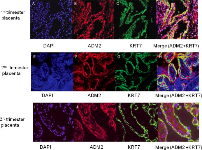
FIG. 3.

Double immunofluorescent staining for ADM2 and cytokeratin 7 in villous trophoblastic tissue, followed by counterstaining with DAPI. (D, H, and L). Double staining for ADM2 and cytokeratin 7 are shown in yellow in first-, second-, and third-trimester placental villi counterstained with DAPI in blue (C, G, and K). Single staining for KRT7 is shown in green in first-, second-, and third-trimester placental villi (B, F, and J). Staining for ADM2 is shown in red in first-, second-, and third-trimester placental villi (A, E, and I). Negative control using rabbit IgG and counterstained with DAPI. Original magnification ×200.
