Figure 4.
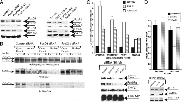
Knockdown of Fox proteins with siRNA. A, T47D cells were treated with 10 nm siRNA to FoxO1, FoxO3a, or FoxO4 (Ambion; left) or with 20 nm smart pool siRNAs for FoxC1, FoxC2, or FoxP1 (Dharmacon; right) for 72 h. Whole-cell lysates prepared and analyzed by SDS-PAGE were blotted and probed with antibodies to FoxO1, FoxO3a, or FoxO4 (left) or with antibodies to FoxP1, FoxC1, and FoxC2. (right). B, T47D cells were transfected with HT-FoxO1 and HT-FoxO3a and simultaneously treated with 10 nm scrambled siRNA, FoxO1 siRNA, or FoxO3a siRNA. The cells were retreated with siRNA at 24 h and incubated for 20 min with 1 μg/ml insulin at 48 h. The plates were washed and nuclear and cytoplasmic extracts were prepared (Materials and Methods). Top row, The extracts were incubated for 30 min with HaloTMR Ligand, and proteins were resolved by SDS-PAGE and visualized using the Typhoon Trio laser scanner. (middle and bottom rows) Separate SDS-PAGE analysis of the same extracts using anti-FoxO1 (middle row) and anti-FoxO3a (bottom row). In this experiment, secondary antibody was infra red (800 nm) tagged and visualized using a Li-Cor Odyssey infrared scanner. C, Effect of knockdown of FoxO3a on PAI-1 promoter activity. T47D cells were treated with 10 nm siRNA for FoxO1, FoxO3a, or with a scrambled siRNA or were left untreated. After 48 h with siRNA, the cultures were retreated with siRNA and transfected with PAI-1-Luc using lipofectamine 2000. The cells were then treated with 1 μg/ml insulin or 1 mm forskolin for 20 h. Luciferase activity was determined and normalized with β-galactosidase. The fold-stimulation by insulin or forskolin was then determined. Top, Luciferase activity; bottom, Fox protein levels were determined by Western blot. D, Treatment of BAE and T47D cells with siRNA for FoxO3a inhibits insulin-increased PAI-1 mRNA. BAE or T47D cells were treated with 10 nm siRNA for FoxO1, FoxO3a, or scrambled siRNA as a control as in panel A. They were re-treated at 48 h, and 1 μg/ml of insulin was added to half of the cultures for an additional 24 h. The cells were lysed and total RNA was prepared and reverse transcribed as described in Materials and Methods. PAI-1 mRNA levels were then determined by RT-qPCR and normalized to GAPDH mRNA levels. The fold stimulation by insulin of PAI-1 mRNA in the cells treated with scrambled, FoxO1, and FoxO3a siRNA in BAE cells (left) and T47D cells (right) is shown. Bottom, Knockdown of FoxO3a in BAE cells was confirmed by Western blotting of whole-cell extracts from BAE cells treated with the different siRNAs (anti-FoxO1, top row; anti-FoxO3a, middle row; and anti-ERK 1 and 2, bottom row).

