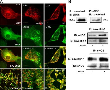
Figure 1.
Insulin induces translocation of CAV-1 and eNOS to the PM and increases the interaction of CAV-1 with eNOS in the membrane. A, Double staining for CAV-1 (red) and eNOS (green). Left column, bAECs cultured in the full medium; center column, serum starved in the basal medium; right column, treated with 10 nm insulin after the serum deprivation. B, The aliquots of membrane extracts were immunoprecipitated followed by Western blots for either eNOS or CAV-1. Top panel, Cultured in full medium; middle and bottom panels, ±10 nm insulin after serum deprivation. Data shown are representative of three to five independent experiments. IB, Immunoblot; IP, immunoprecipitation; KD, kilodalton.

