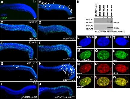
Figure 5.
Udu dynamics and localization during DNA repair and replication. (A and B) Immunohistochemical staining of γ-H2AX in nonirradiated embryos at 22 hpf embryos: γ-H2AX foci in wild-type embryos (A) and udutu24 embryos (B). (C–H) Wild-type and udutu24 embryos were irradiated with 15 min of UV and stained with γ-H2AX at different time after irradiation. (C and D) γ-H2AX staining after 1 h post-UV treatment. Both UV-treated wild-type and udutu24 embryos showed an increased in γ-H2AX foci. Hence, in the presence of extrinsic DNA damage agents, udutu24 mutants were able to response accordingly by recruiting DNA repair proteins via the activation of γ-H2AX as indicated by the amplified fluorescence staining. (E and F) γ-H2AX staining of embryos 5 h post-UV irradiation. The level of DNA damage after 5 h of repair were similar in both wild-type and udutu24 embryos. (G and H) The number of cells with γ-H2AX signals was significantly reduced in the trunk and tail regions of wild-type and udutu24 embryos after 31 h post-UV irradiation. (I and J) γ-H2AX staining of wild-type and udutu24 embryos injected with p53-MO. White arrows indicate the γ-H2AX foci. (K) Coimmunoprecipitation of MYC-tagged MCM proteins and FLAG-tagged PAH-L+SANT-L domains. FLAG-tagged PAH-L+SANT-L is immunoprecipitated from the cell lysates and detected with anti-FLAG antibody as a control and with anti-MYC antibody to detect the interacting proteins, MCM3 and MCM4, shown in lanes 4 and 5. (L–O) PAH-L and SANT-L domains are associated with replication foci during S phase. COS7 cells were synchronized at the G1/S border with aphidicolin, released into S phase, labeled with BrdU and fixed at different time points and submitted to laser scanning confocal microscopy for detection: DAPI (blue) (L and P), PAH-L+SANT-L (green) (M and Q), BrdU (red) (N and R), and merged images (yellow) (O and S) show the pattern of replication in early S phase (L–O) and late S phase (P–S). (T–W) Localization of Udu in pericentromeric heterochromatin: DAPI (blue) (T), Udu (green) (U), HP1β (red) (V), and merged image (yellow) (W) shows that Udu is colocalized with HP1β, a protein marker for pericentromeric heterochromatin. IB, immunoblotting; IP, immunoprecipitation.

