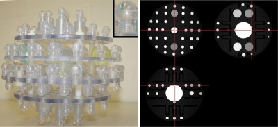
Figure 1.
ADNI phantom. A photograph of the internal components of the ADNI phantom is shown. Each of the spheres is filled with a copper sulfate solution. The colored spheres contain differing solution concentrations. The small inset provides a detailed view of a single sphere and postcomponent. A triplanar view of a phantom image acquired with the MP-RAGE used in the ADNI protocol is also shown.

