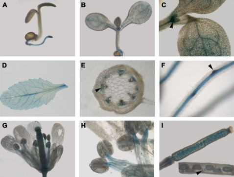
Fig. 8.
Organ and tissue expression of pHKL1-GUS. (A) Seedlings grown for 3 d on MS plates. (B) Seedlings grown for 7 d on MS plates. (C) Shoot of a 5-d-old seedling, with the arrowhead pointing to specific stain in the meristem. (D) Leaf from a 21-d-old plant. (E) Stem cross-section, with the arrowhead pointing to staining of phloem. (F) Root of a 10-d-old seedling, with the arrowhead pointing to enhanced staining at the site of lateral root initiation. (G) Opened flower. (H) Anthers and filaments. (I) Developing silique, with insert showing a mature silique and an arrowhead pointing to the funiculus of a developing seed.

