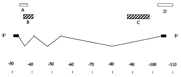
Figure 2. Probes for the Ubx Transcription Unit.

The positions of the four Ubx exons are indicated relative to a scale in kb. Only one of the alternative splicing patterns is indicated. The major mature transcripts of 3.2 and 4.3 kb, while differing in their inclusion of the internal exons, are derived from a roughly 77 kb primary transcript. The boxes above indicate the positions of probes used in this study. Unless indicated otherwise, probes described as 5′ and 3′ refer to probes B and C, respectively. Probe B is recessed from the 5′ end by 3.5 kb, and probe C falls about 10 kb short of the 3′ end. The 5′ ends of these probes are separated by 52 kb. This schematic of the Ubx transcription unit is based on O'Connor et al. (1988).
