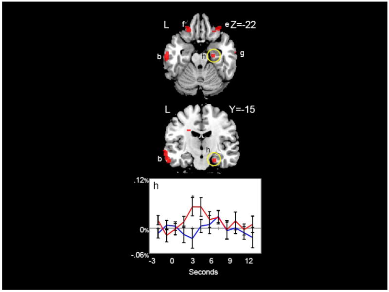
Figure 5.

Temporal, Frontal, and Parahippocampal Regions. Regions in which content words elicited significantly greater activation than function words (red) are shown. Hemodynamic time courses to content (red) and function (blue) words in the right parahippocampal gyrus (h) are shown. Time points with significant differences between content and function words are indicated with an asterisk. Coordinates are in MNI space.
