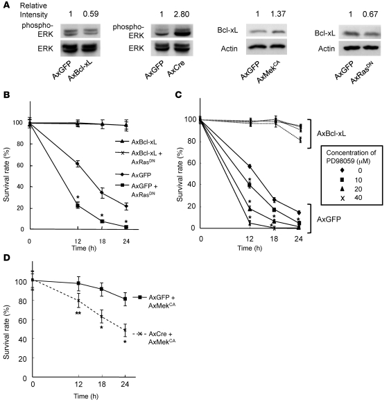
Figure 5. Ras-Mek-Erk pathways are upstream of Bcl-xL in osteoclasts.
(A) Bcl-xfl/fl osteoclasts were infected with AxGFP, AxCre, AxBcl-xL, AxMekCA, or AxRasDN. Bcl-xL overexpression by AxBcl-xL infection suppressed, and knockout of Bcl-x gene by AxCre infection increased, Erk activity, as determined by the amount of phospho-Erk in Bcl-xfl/fl osteoclasts. In contrast, AxMekCA infection increased, and AxRasDN infection decreased, Bcl-xL expression. Relative intensity of the bands on each gel, measured by densitometry, is shown above each lane. (B) Bcl-xfl/fl osteoclasts were infected with AxGFP, AxGFP plus AxRasDN, AxBcl-xL, or AxBcl-xL plus AxRasDN. Reduced osteoclast survival by RasDN overexpression was completely rescued by Bcl-xL overexpression. *P < 0.01 versus AxGFP-infected cells. (C) Bcl-xfl/fl osteoclasts were infected with AxGFP or AxBcl-xL, and then treated with the indicated concentrations of MEK inhibitor PD98059. PD98059 treatment dose-dependently suppressed the survival of osteoclasts, which was completely rescued by Bcl-xL overexpression. *P < 0.01 versus untreated osteoclasts. (D) Bcl-xfl/fl osteoclasts were infected with AxGFP or AxCre together with AxMekCA. Prosurvival effect of MekCA overexpression was partially suppressed by Bcl-x deletion. *P < 0.01, **P < 0.05 versus AxGFP-AxMekCA–infected cells. All results are mean ± SD of 6 cultures.

