Fig. 4.
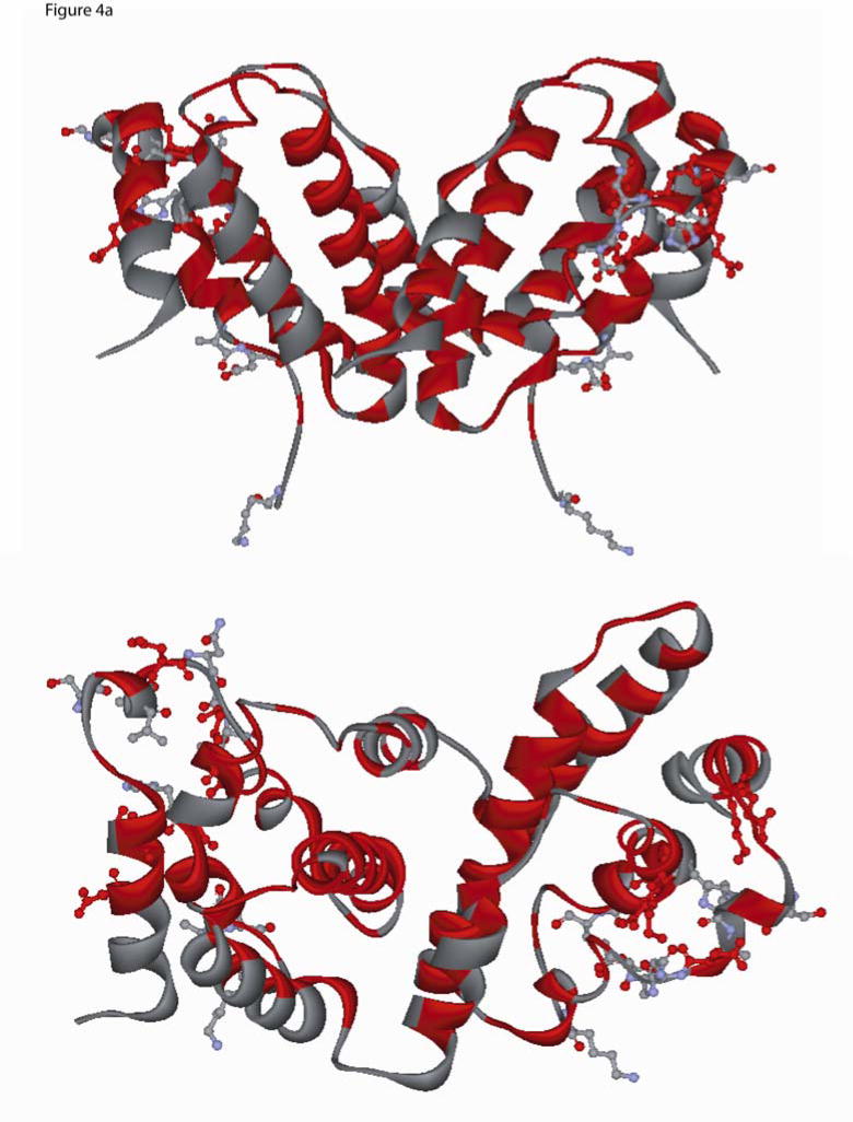
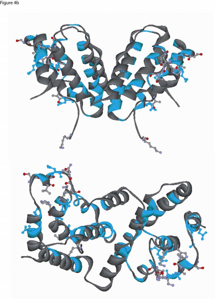
Two different ribbon view of the interferon-gamma dimer (PDB ID, 1FG9) (a) The location of the conserved residues identified from mammalian-only sequence alignment are shown in red (see Fig. 3a), and the residues that are within 3.5 Å from IFN-γ receptor atoms are shown in ball-and-stick and colored by element type (Carbon:light grey, Oxygen:red, Hydrogen:white, Nitrogen:light blue). Residues that belong to the mammalian conserved set and are present within 3.5 Å are shown in red color ball-and-stick (upper panel is top view and lower panel is side view (non stereo)). (b) Conserved residues based on the frog, chicken, human and fish sequence alignment were painted Cyan (Fig 3a), and the residues that are present within 3.5 Å from the IFN-γ receptor are shown in ball-and-stick colored by element type (Carbon:light grey, Oxygen:red, Hydrogen:white, Nitrogen:light blue). Residues which belong to the conserved set and are present within a 3.5 Å are shown in cyan color ball-and-stick (upper panel is top view and lower panel is side view (non stereo)).


