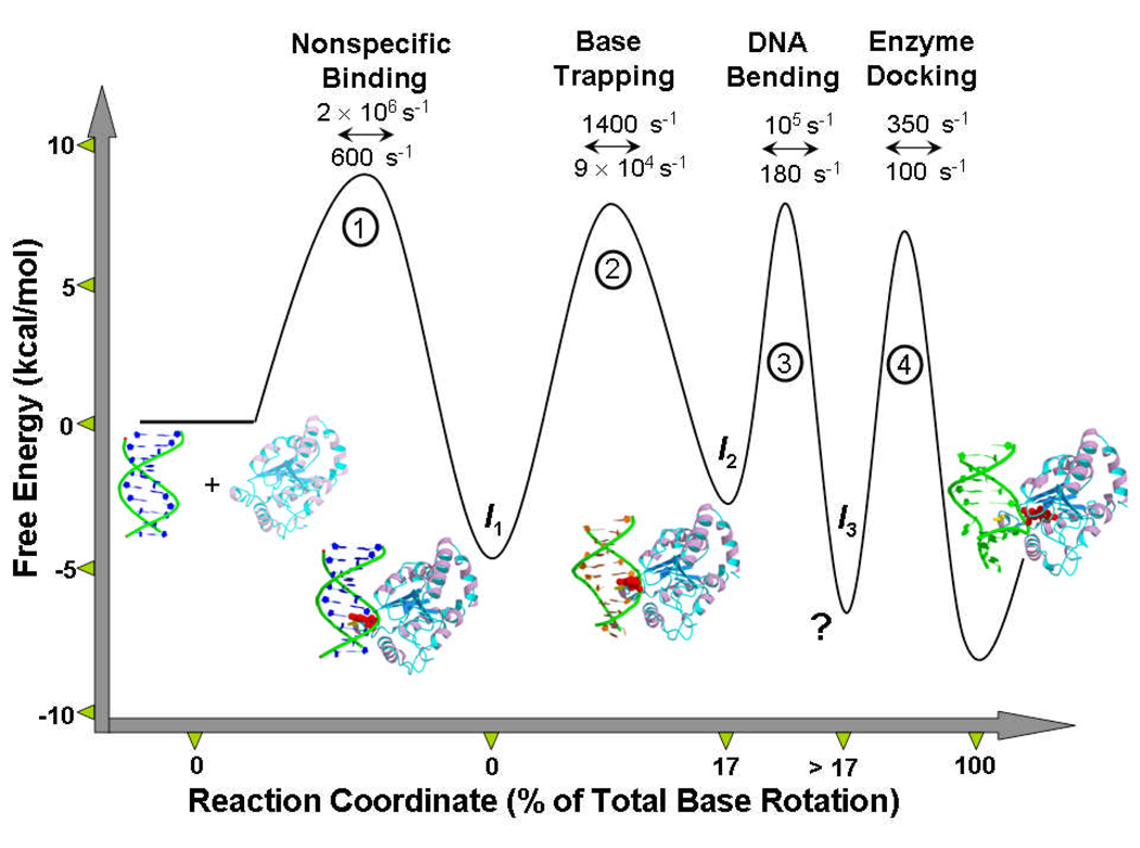
Figure 6.
The reaction coordinate for uracil flipping by UNG. The microscopic rate constants have been calculated by combining NMR[19,34] and rapid kinetic measurements[24,25]. The profile pertains to 25 °C. The structures are: free human UNG (pdb 1AKZ), intermediate 1 (encounter complex with B DNA, model)[23], intermediate 2 (partially flipped intermediate state, pdb 2OXM), intermediate 3 (detected kinetically, no structural model)[20,25], final flipped state (pdb 1EMH)[33]. Since the rates by neccesity were obtained using different substrates and by extrapolation of the base pair opening rates to 25 °C, the values should only be considered best approximations.

