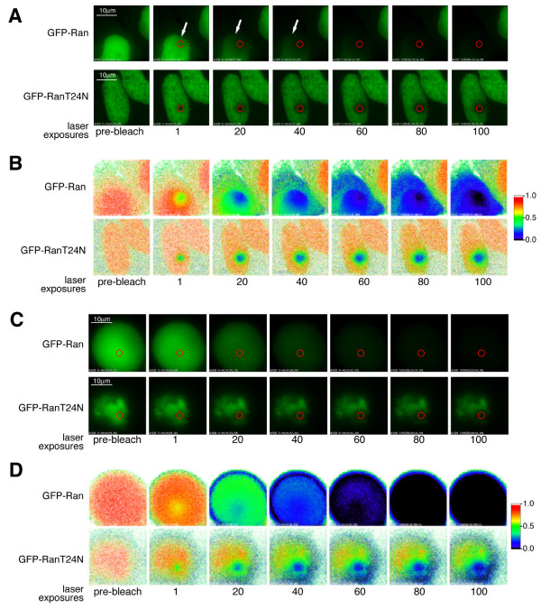
Figure 6.
FLIP analysis of GFP-RanWT and GFP-RanT24N. Successive images of individual cells expressing GFP-RanWT or GFP-RanT24N in interphase (A) or mitosis (C) after photobleaching within a defined spot within the nucleus for the number of exposures shown. (B,D) False-colour imaging showing the relative change in fluorescence for the cells shown in (A) and (C), from orange through green to blue for increasing change, illustrating bleaching of the fluorescence signal.

