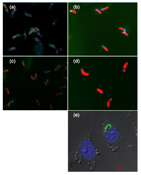
Figure 2.
IFAT analysis demonstrating specific binding of scFv AB28 to sporozoites of E. tenella (a) and lack of interaction with sporozoites of E. acervulina (b) and E. brunetti (d). Negative control (binding of irrelevant scFv) is shown in panel (c). Blue fluorescence (DAPI), staining of nuclei; red fluorescence (Evans blue), staining of sporozoite refractile bodies and cytoplasm; green fluorescence (FITC), specific antibody staining. In panel (e), a fluorescence microscopy image is shown of intracellular sporozoite in infected HepG2 cell. Staining was performed with scFv AB28 followed by anti-c-myc MAb and Alexa488-conjugated anti-mouse antibody.

