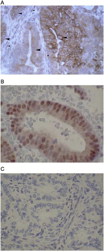
Fig. 6.
Detail of PRB staining and nuclear exclusion at high magnification. (A) PRB is cytoplasmic in some poorly differentiated tumors. Note the central blue staining of the nucleus of the tumor cells (large arrowheads), with surrounding brown staining in the cytoplasm. In comparison, the stromal cells surrounding the tumor demonstrate brown nuclear staining for PRB (small arrowheads). (B) PRB retains nuclear staining in some well-differentiated endometrial tumors. (C) Negative control for IHC staining. (For interpretation of the references to colour in this figure legend, the reader is referred to the web version of this article).

