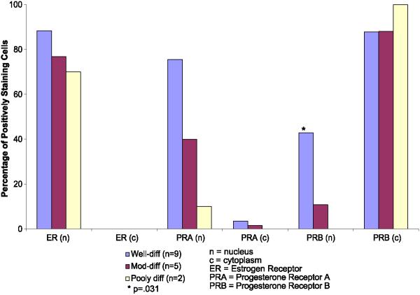
Fig. 7.
Percentage of tumor cells demonstrating positive staining for nuclear and cytoplasmic receptors in well-, moderately- and poorly-differentiated endometrial cancers. ER = estrogen receptors and PRB = progesterone B receptors. Blue bars = well-differentiated tumors (n = 9), red bars = moderately differentiated tumors (n = 5), and beige bars = poorly differentiated tumors (n = 2). *The presence of PRB in the nucleus is significantly correlated with tumor differentiation using the Mantel-Haenszel test, P = 0.031. (For interpretation of the references to colour in this figure legend, the reader is referred to the web version of this article).

