Figure 1.
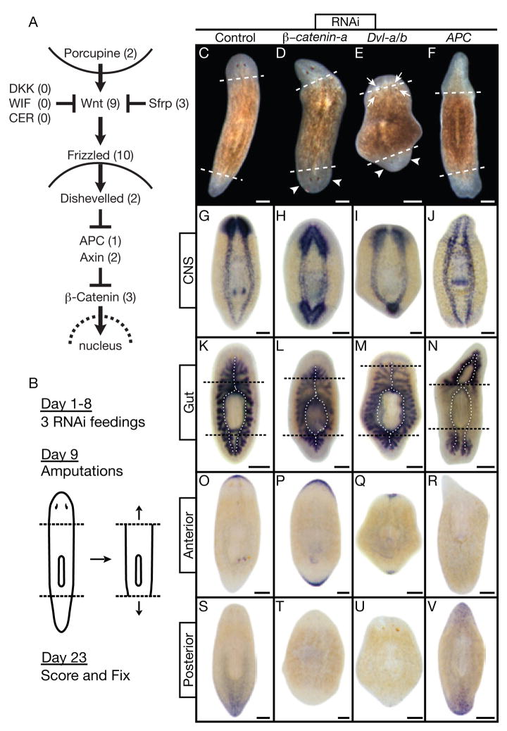
Signaling through β-catenin defines head vs. tail during regeneration. (A) Canonical Wnt pathway. Numbers: S. mediterranea homologs identified and silenced. (B) Experimental strategy. (C through V) Trunk fragments 14 days post-amputation. Dashed lines: amputation planes. Control: unc-22(RNAi). (C through F) Live animals. (D, E) Smedβ-catenin-a(RNAi) and SmedDvl-a/b(RNAi) posterior blastemas formed heads with photoreceptors (arrowheads). SmedDvl-a/b(RNAi) fragments also developed ectopic photoreceptors, often within old tissues (arrows). (F) SmedAPC(RNAi) anterior blastemas formed tails. (G through V) In situ hybridizations, n≥5 per marker. Markers: CNS, pro-hormone convertase 2 (PC2); Gut, Porcupine (SmedPorcn-a); Anterior, secreted Frizzled-related protein (SmedSfrp-a); Posterior, Frizzled (SmedFz-d). (G,K) Normal brain and ventral nerve cords; single anterior and dual posterior gut branches (dotted lines). (H,I,L,M) Smedβ-catenin-a(RNAi) and SmedDvl-a/b(RNAi) posterior blastemas developed brain tissue and head-like gut branches. (J,N) SmedAPC(RNAi) anterior blastemas developed tail-like nerve cords and gut branches. (O,S) Normal anterior and posterior marker expression. (P,Q,T,U) Smedβ-catenin-a(RNAi) and SmedDvl-a/b(RNAi) induced anterior marker expression at both ends while the posterior marker was virtually undetectable. (R,V) SmedAPC(RNAi) induced posterior marker expression at both ends while the anterior marker was virtually undetectable. Scale bars: 200 μm.

