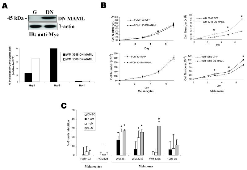
Figure 2. Inhibition of Notch signaling suppresses melanoma, but not melanocyte growth in vitro.
A, Expression of DN-MAML protein in melanoma cell line WM 3248 was detected by immunoblotting for the myc-tagged DN-MAML. Effect of DN-MAML expression on Notch1 targets Hes1, Hey1 and Hey2 was determined by quantitative RT-PCR in 2 melanoma cell lines, WM3248 and WM1366.
B, 7 day growth curves of melanocytes and melanoma cells infected with DN-MAML or GFP lentiviruses. * p<0.05 (Student's T-test).
C, Growth inhibition in melanocytes and melanoma cells in the presence of increasing concentrations of a γ-secretase inhibitor. Cell growth was determined by MTT analysis. Results are percentage of growth inhibition compared with untreated controls (adjusted to 0%). * p<0.005 (Student's t-test).

