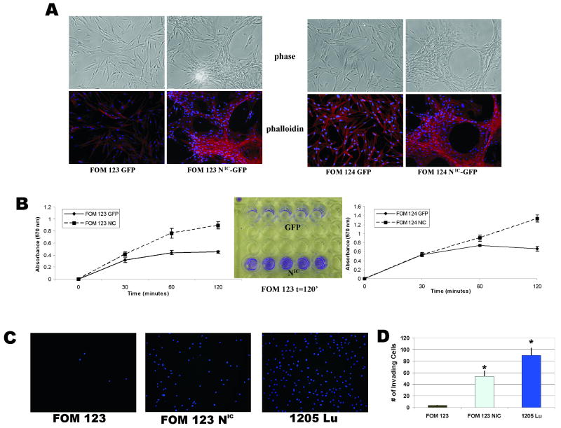
Figure 4. Constitutive Notch1 activation induces cytoskeletal, adhesion, and migratory changes in primary melanocytes.
A, Phalloidin immunostaining reveals morphological changes induced by active Notch1 expression. FOM 123 and 124 NIC cells form networks of capillary like structures when grown in monolayer as compared to the normal dendritic growth pattern of normal GFP-infected cells.
B, Cell adhesion assay demonstrated that NIC-infected FOM cells possess increased adhesive properties compared to GFP-infected controls within 2 hours post-plating. *p<0.005 (Student's t-test).
C & D, NIC- and GFP-infected melanocytes were subject to Boyden chamber assays to test their ability to migrate through matrigel. After 24 hours, membranes stained with DAPI demonstrated enhanced ability of NIC-infected FOM cells to migrate; 1205Lu melanoma cells acted as a positive control.

