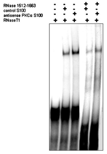
Figure 3. Assessment of the binding of cytoplasmic factors in PKCα-depleted cells to sequences on the 3′ UTR of LPL.

Gel shift and RNase protection analyses have been performed to assess the binding of cytoplasmic factors to sequences on the LPL 3′ UTR. [32P]UTP-labelled transcripts corresponding to nt 1512–1663 of LPL mRNA were incubated with 1–5 μg of S-100 extract from control and antisense PKCα-treated adipocytes, followed by the digestion of unprotected RNA with RNase T1. A specific RNaseT1 protected gel shift was obtained consistently only with the transcript 1512–1663 as is illustrated in the Figure. The control cell extract from cells treated with Lipofectin® alone provided a weaker gel shift.
