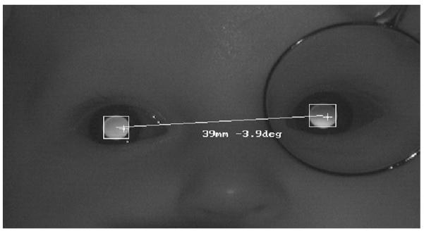
FIGURE 1.

An example PowerRefractor image from an infant. The distribution of light in the pupils provides simultaneous estimates of defocus in the two eyes, and the positions of the Purkinje images relative to the center of the pupils provide an estimate of gaze position. The image also demonstrates the technique used in this study for the relative validation. A positive-powered lens was held before one eye, producing a myopic crescent in the pupil, and the induced anisometropia was determined as a function of lens power. (The camera aperture size was kept in the recommended range of 5.6–8 throughout data collection.)
