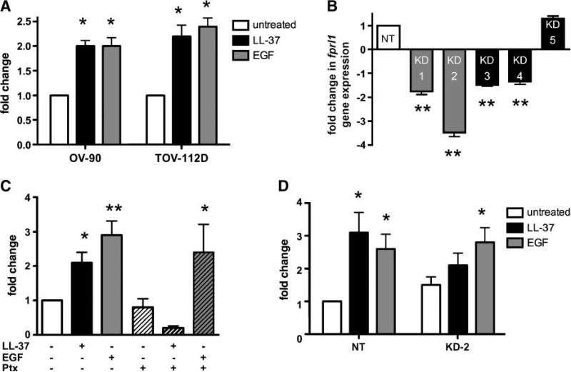
FIGURE 3.
LL-37 mediates ovarian cancer cell migration and invasion through FPRL1. A. Graphic representation of ovarian cancer cell invasion. Serum-starved cells were seeded onto Matrigel-coated inserts in medium containing 10 μg/mL of LL-37 or 10 ng/mL of EGF. Columns, mean fold change of the mean fluorescence intensity of invaded cells compared with unstimulated controls; bars, SE (n = 3). B. Graph depicting fprl1 gene expression in knockdown (KD) cells. SK-OV-3 cells stably transduced with lentiviruses containing FPRL1-specific shRNA (KD-1 to KD-5) or nontarget (NT) sequences were analyzed by qPCR. Columns, mean of three independent experiments compared with nontarget cells; bars, SE. C. Graphic representation of SK-OV-3 ovarian cancer cell invasion through Matrigel-coated inserts. Untreated and Ptx-treated SK-OV-3 cells were stimulated with LL-37 or EGF as described above. P values for LL-37 or EGF groups were determined from their respective untreated or Ptx-treated alone controls. D. Graphic representation of SK-OV-3 nontarget cells and FPRL1 KD-2 cell invasion stimulated with LL-37 or EGF as above (*, P < 0.05; **, P < 0.01).

