
Fig. 6.
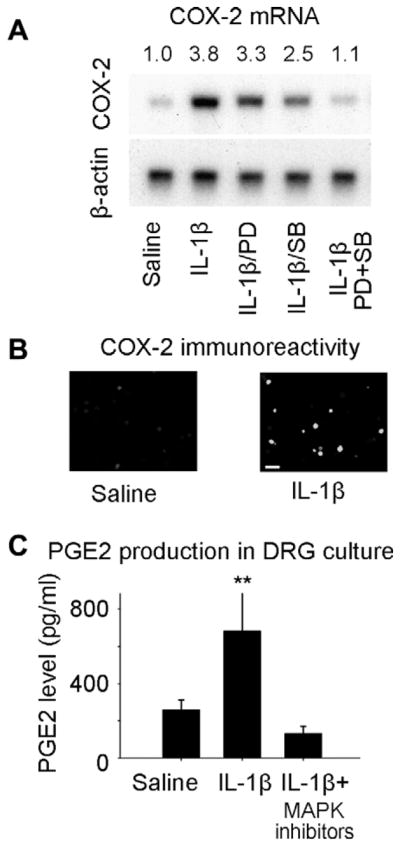
(A) Signal pathways involved in COX-2 induction in DRG neurons cultured from na rats 6 h after exposure to IL-1β. (A) Application of IL-1β to cultured DRG neurons produced an increase in COX-2 mRNA that was partially blocked by administration either of SB203580, a p38 MAP kinase inhibitor or of PD98059, an ERK kinase inhibitor. Co-application of the p38/ERK inhibitors blocked COX-2 induction almost completely. The COX-2/β-actin signal density ratio is presented above the image. (B) COX-2 immunoreactivity in cultured DRG neurons after IL-1β. Scale bar = 50 μm (C) Prostaglandin levels in the media of cultured DRG neurons were increased by IL-1β in a p38/ERK dependent fashion.