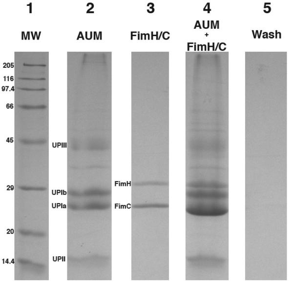
Figure 1. Gel electrophoresis (SDS-PAGE) analysis of FimH binding to mouse urothelial plaques.
Purified mouse urothelial plaques were incubated with FimH/C and washed excessively; the proteins in the complex were resolved by SDS-PAGE and stained with Coomassie blue. Lane 1: Molecular weight marks. Lane 2: Mouse urothelial plaques (AUM). The positions of the 4 uroplakins are indicated on the left. Lane 3: Recombinant FimH/C complex. Lane 4: Urothelial plaques incubated with FimH/C and washed 3 times with buffers showing FimH/C was complexed with urothelial plaques. Note that FimH is clearly visible while FimC co-migrates with the UP Ia band. Lane 5: Supernatant of the last wash.

