
Figure 4. FimH binding-induced movements of the transmembrane helices of uroplakins.
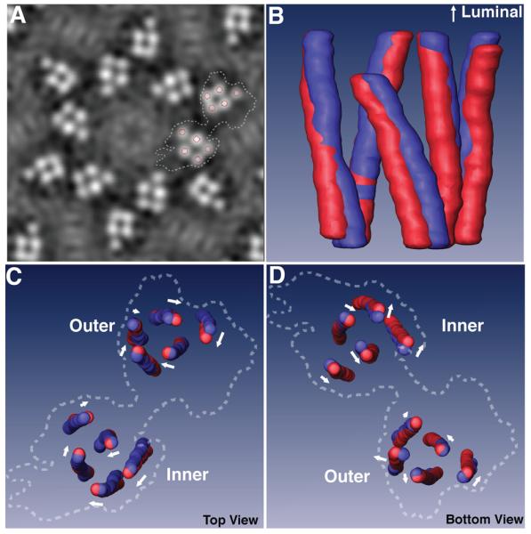
(A) A slice of the density map in the middle of the transmembrane domain of the FimH-bound 16-nm particle. Circles mark the center of the transmembrane helices in the segmentation process, and dashed lines outline a subunit. (B) Side view of the superimposed, segmented transmembrane helices of the inner subdomains of the native (blue) and FimH-bound (red) particles. (C) and (D) Superimpositions of the segmented transmembrane helices of the inner and outer subdomains viewed from the luminal (C) and the cytoplasmic (D) sides. The arrow near each helix approximates the direction and magnitude of the helix's movement induced by FimH-binding. Dashed lines again indicate the outlines of a whole subunit.