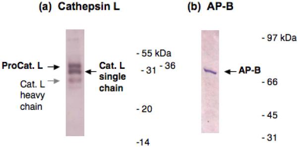
Figure 6. Cathepsin L and aminopeptidase B (AP-B) in AtT-20 cells illustrated by western blots.

Western blots of cathepsin L (panel a) and AP-B (panel b) were conducted in AtT-20 cells. The presence of cathepsin L and AP-B was demonstrated by western blots with specific antisera to each protease, performed as described in the methods.
(a) Cathepsin L in AtT-20 cells. Cathepsin L bands observed by anti-cathepsin L western blot correspond to procathepsin L (∼38-40 kDa, black arrow), the mature single chain form of cathepsin L (∼28 kDa, black arrow), and a light band corresponding to the heavy chain form of cathepsin L (∼24 kDa, gray arrow) [24].
(b) AP-B in AtT-20 cells. The AP-B visualized by western blot with anti-AP-B corresponds to a band of ∼72 kDa [19].
