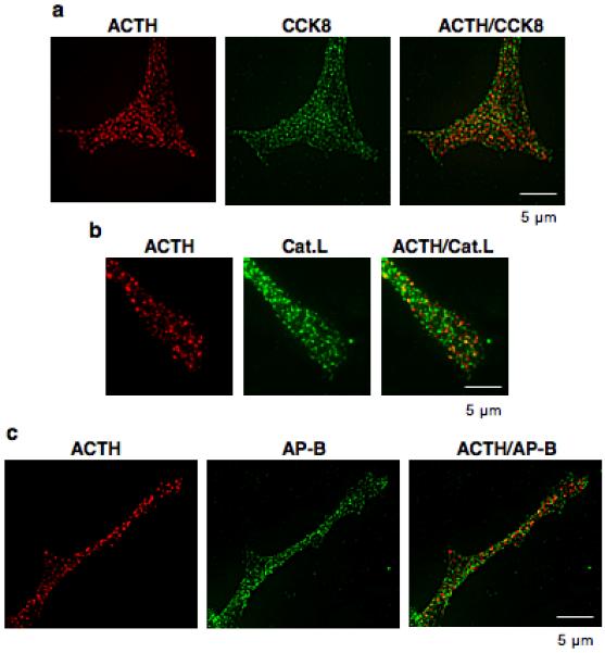
Figure 9. Colocalization of ACTH in secretory vesicles with CCK8, cathepsin L, and aminopeptidase B in neuritic extensions of AtT-20 cells.

The colocalization of ACTH with CCK8, cathepsin L, and aminopeptidase (AP-B) was examined in neuritic-like extensions of AtT-20 cells which contain secretory vesicles. Panel ‘a’ shows the colocalization of ACTH (red fluorescence) with CCK8 (green fluorescence) shown by the merged yellow fluorescence. Panels ‘b’ and ‘c’ show the colocalization of ACTH (red fluorescence) with cathepsin L and aminopeptidase B (green fluorescence) indicated by the yellow fluorescence in the merged images.
