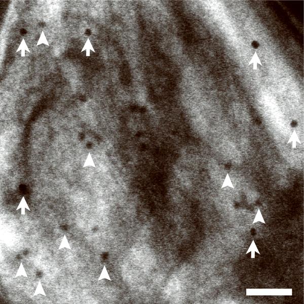
Figure 2.
Bright-field STEM tomography of 1 μm-thick C. reinhardtii. (a) 2D projection image of entire C. reinhardtii in the xy plane. (b) 25-nm thick slices across the xy (top panel) and xz (bottom panel) planes of BF STEM tomogram. (c) 10-nm thick slice across tomogram recorded from the region marked in a. (d) Expanded areas from tomogram in c showing fine ultrastructural details. Left panel: the two faces of a vesicle membrane are resolved indicating a resolution within 5 nm (arrows). Right panel: membranes of thylakoid stacks. Scale bars, 1 μm (a,b), 500 nm (c), 50 nm (d).

