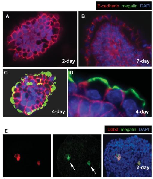
FIG. 1.
Distribution of E-cadherin and megalin proteins in embryoid bodies. Embryoid bodies formed from the aggregation of mouse wild-type RW4 ES cells were harvested in various stages, fixed, sectioned, and subjected to immunofluorescence analysis. Merged images of indirect immunofluorescence of E-cadherin (red), megalin (green), and nuclei labeled by DAPI (blue) are shown (A—D): (A) A representative 2-day-old spheroid prior to the formation of a surface primitive endoderm layer. B: A more mature, 7-day spheroid furnished with a primitive endoderm layer. C: A representative 4-day-old spheroid exhibiting a superficial primitive endoderm layer, and (D) a higher magnification to show the mutually exclusive distribution of megalin and cadherin in surface endoderm cells. Mouse wild-type RW4 ES cells were used to make these spheroids. E: A 2-day-old spheroid prior to the formation of a surface primitive endoderm layer contains Dab2 (red)- and megalin (green)-positive primitive endoderm cells (arrows) in the interior of the spheroids. [Color figure can be viewed in the online issue, which is available at www.interscience.wiley.com.]

