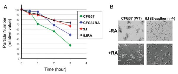
FIG. 3.
Cell adhesion of differentiated and undifferentiated wild-type and E-cadherin null ES cells. A: Prior differentiated (4 days with retinoic acid) and undifferentiated wild-type and E-cadherin null ES cells were assayed for cell adhesion by a reduction of particle number (cells and aggregates) over a 3-h period using a Coulter counter. CFG37, undifferentiated wild-type ES cells; CFG37RA, retinoic acid-differentiated CFG37 ES cells; 9J, E-cadherin null ES cells; 9JRA, retinoic acid-differentiated 9J ES cells. The standard error of the mean from the three readings of particle number at each time point of a sample is indicated by an error bar, which is typically smaller than 10%. B: Cell morphology shown by phase contrast microscopy of wild type or 9j (E-cadherin null) ES cells, with or without prior differentiation with 1 μM retinoic acid for 4 days. The cells were first dispersed as suspension of individual cells using trypsin and EDTA. Then, equal numbers of the cells were suspended in calcium-containing culturing medium and kept in gentle motion in a 37°C incubator for 4 h. Morphology and patterns of cell aggregations were shown by phase contrast microscopy. [Color figure can be viewed in the online issue, which is available at www.interscience.wiley.com.]

