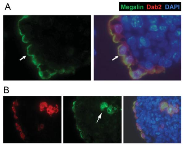
FIG. 4.
Apical polarity of surface endoderm cells in E-cadherin null embryoid bodies. A: A representative seven-day spheroid produced from the aggregation of 9j E-cadherin-null ES cells is shown. The Dab2-positive endoderm cells formed a layer lining the surface and are stained positive for megalin on the apical surface (arrow). Megalin (green) and the merged images of indirect immunofluorescence of Dab2 (red), megalin (green), and nuclei labeled by DAPI (blue) are shown. B: An E-cadherin (-/-) embryoid body contains Dab2- and megalin-positive primitive endoderm cells located in both the surface layer and the interior (arrow) of the spheroids. Note that megalin is polarized in surface cells but not in the inner cells. [Color figure can be viewed in the online issue, which is available at www.interscience.wiley.com.]

