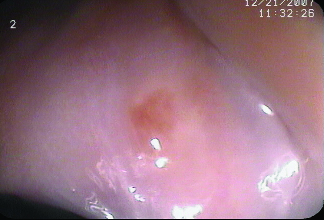
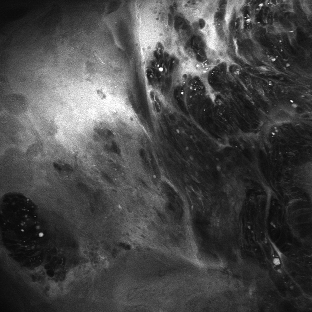
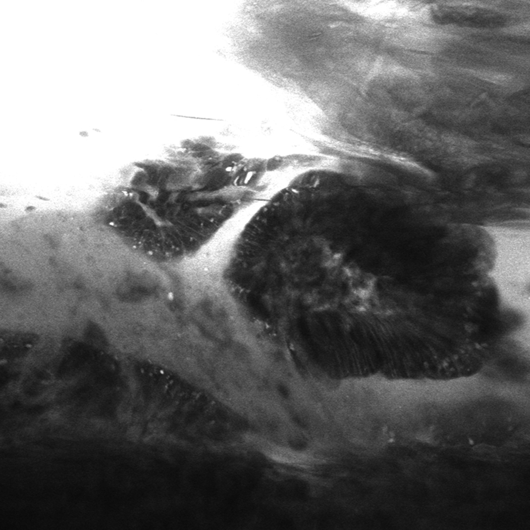
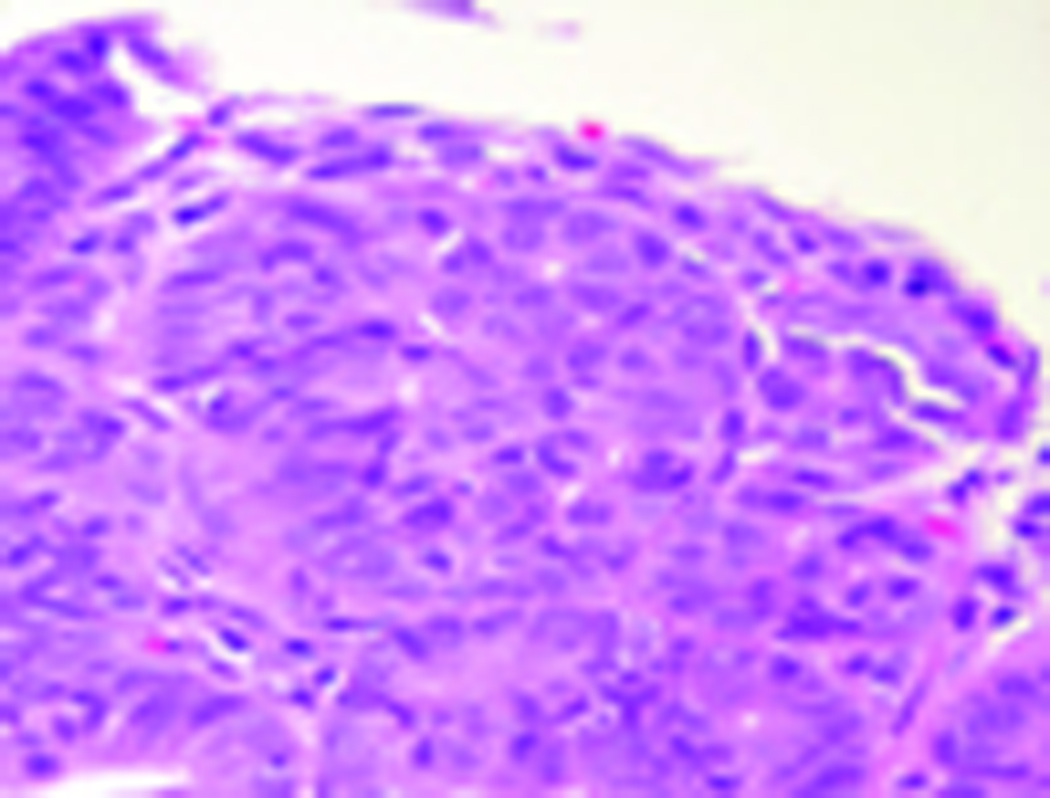
Figure 3.
(A) Unmagnified standard white light endoscopic image of a tiny island of Barrett’s esophagus obtained with the endomicroscope prior to endomicroscopic imaging. (B and C) Confocal endomicroscopy images of the island shows glands with irregularly-shaped, distorted dark cells, indistinct cell borders, and loss of normal crypt architecture suggestive of high grade dysplasia (D) Histopathology confirmed Barrett’s esophagus with HGD in the endoscopic mucosal resection specimen


