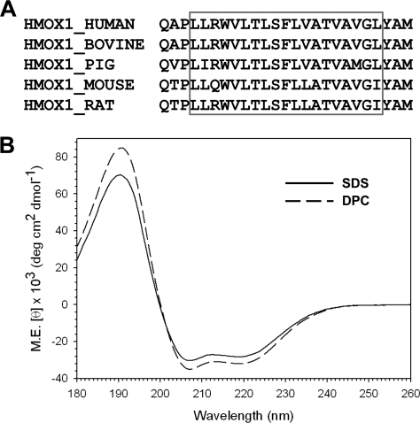
FIGURE 3.
The CD spectra of the human HO-1 TMS. A, sequence alignment of the last 25 amino acids in the C-terminal of vertebrate HO-1s. The red box indicates the predicted TM α-helix. B, CD spectra of a 20 μm solution of synthetic human HO-1 C-terminal 25 residue peptide in 10 mm SDS (solid line) or DPC (dashed line) at 25 °C in 20 mm phosphate buffer (pH 7.0).

