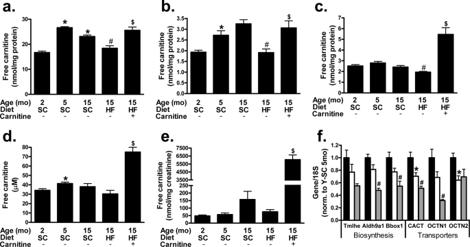
FIGURE 2.
Whole body carnitine status is compromised by lifelong high fat feeding and restored by carnitine supplementation. Tandem MS/MS was used to measure free carnitine levels in mixed gastrocnemius (a), liver (b), kidney (c), plasma (d), and urine (e) harvested 4 h after food withdrawal from young (Y, 5 month) or old (O, 15 month) animals fed a standard (SC) or high fat (HF) diet. Quantitative reverse transcription-PCR was used to measure the expression of hepatic genes involved in cellular (OCTN2) and mitochondrial (CACT and OCTN1) carnitine transport, as well as genes encoding the first (Trimethyllysine hydroxylase-ϵ (Tmlhe)), third (aldehyde dehydrogenase-9 family, member A1 (Aldh9a1)), and fourth/final (γ-butyrobetaine hydroxylase-1 (Bbox1)) reactions involved in carnitine biosynthesis; young (black), old (white), and old-HF (gray) (f). Data represent means ± S.E. from 5–8 animals per group. A one-way analysis of variance with Tukey's post hoc test was used to examine the effects of aging, whereas a Student's t test was used to determine differences due to HF diet and carnitine supplementation. *, p < 0.05 due to aging; #, p < 0.05 due to HFD; $, p < 0.05 due to carnitine supplementation.

