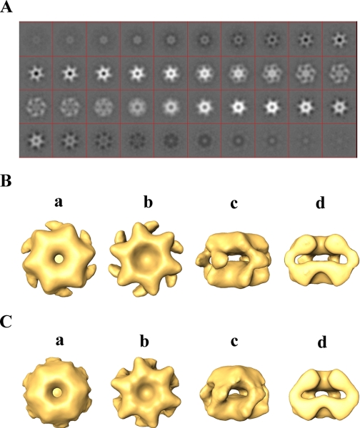
FIGURE 1.
Three-dimensional reconstruction of PAN in the presence of ATPγS. A, serial successive, 0.4-nm-thick slices through the three-dimensional-reconstructed volume of PAN (top ring to bottom ring) in the presence of ATPγS. B, orthogonal views of reconstructed PAN in the presence of ATPγS. a and b, top and bottom views, respectively; c, side view; d, cut-away side view. C, a surface rendering view of the three-dimensional reconstruction of PAN-ΔN (1–73)-ATPγS complex. a and b, top and bottom views, respectively; c, side view; d, cut-away side view.

