FIGURE 3.
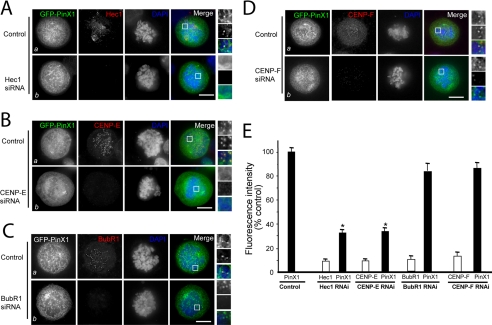
Kinetochore localization of PinX1 requires Hec1 and CENP-E. A, Hec1 determines the kinetochore localization of PinX1. Aliquots of HeLa cells were transfected with oligonucleotides (control and siRNA for Hec1) and GFP-PinX1 for 48 h, followed by fixation and immunocytochemical staining as described under “Experimental Procedures.” Optical images were collected from HeLa cells transfected with control siRNA (Control panel) and Hec1 siRNA (Hec1 siRNA panel). Suppression of Hec1 eliminates the kinetochore localization of PinX1, although its perichromosomal distribution is not altered. Scale bars, 10 μm. B, CENP-E determines the kinetochore localization of PinX1. Aliquots of HeLa cells were transfected with oligonucleotides (control and siRNA for CENP-E) and GFP-PinX1 for 48 h, followed by fixation and immunocytochemical staining as described above. Optical images were collected from HeLa cells transfected with control siRNA (Control panel) and CENP-E siRNA (CENP-E siRNA panel). Suppression of Hec1 eliminates the kinetochore localization of PinX1, although its perichromosomal distribution is not altered. Scale bars, 10 μm. C, kinetochore localization of PinX1 is independent of BubR1 in the kinetochore. Aliquots of HeLa cells were transfected with oligonucleotides (control and siRNA for BubR1) and GFP-PinX1 for 48 h, followed by fixation and immunocytochemical staining as described above. Optical images were collected from HeLa cells transfected with control siRNA (Control panel) and BubR1 siRNA (BubR1 siRNA panel). Suppression of BubR1 does not alter the kinetochore localization of PinX1 and PinX1 associated with perichromosome regions. Scale bars, 10 μm. D, kinetochore localization of PinX1 is independent of Cenp-F in the kinetochore. Aliquots of HeLa cells were transfected with oligonucleotides (control and siRNA for Cenp-F) and GFP-PinX1 for 48 h, followed by fixation and immunocytochemical staining as described above. Optical images were collected from HeLa cells transfected with control siRNA (Control panel) and Cenp-F siRNA (Cenp-F siRNA panel). Suppression of Cenp-F does not alter the kinetochore localization of PinX1 and PinX1 associated with perichromosome regions. Scale bars, 10 μm. E, quantitation of Hec1, CENP-E, BubR1, and Cenp-F levels at kinetochores of control and siRNA-treated cells. The pixel intensities of Hec1, CENP-E, Hec1, and Cenp-F (normalized to the ACA signal) in control (closed bars) and Hec1-repressed, CENP-E-repressed, BubR1-repressed, and Cenp-F-repressed cells were measured. Values represent the means ± S.E. of at least 100 kinetochores in 11 different cells. The intensities of target proteins are expressed as open bars, although PinX1 intensities are marked by closed bars. *, p < 0.01 compared with that of the control.

