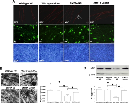
FIGURE 7.
Down-regulation of P2X7 receptor expression in CMT1A SC improves myelination. Wild type and CMT1A SC from newborn rats were electroporated in the presence of negative control shRNA or in the presence of a shRNA plasmid targeting P2X7 (wild type shRNA and CMT1A shRNA). SC were then seeded onto DRG neurons and allowed to myelinate for up to 4 weeks. A, immunocytochemistry for MBP on DRG/SC co-cultures. Axons are stained with an anti-neurofilament antibody, and nuclei of SC are stained with 4′,6-diamidino-2-phenylindole (DAPI). Co-cultures with NC-transfected CMT1A SC show axonal segments devoid of myelin (arrows), along with normally myelinated internodes. Co-cultures with shRNA-P2X7-transfected SC show a higher extent of myelination. B, light microscopy morphometric analysis of myelination in DRG/SC co-cultures. Upper panels show co-cultures stained with Sudan black. Arrows indicate demyelinated internodes. The histograms show the density of myelin segments in the various co-cultures. Data are mean numbers of myelinated segments/mm2 ± S.E. in 250–300 fields of four independent cultures for each condition. C, Western blot analysis of MPZ levels in lysates of the various co-cultures. The histograms show the densitometric quantification of band intensity: values shown are the mean ± S.E. of three independent experiments. β-TUB, β-tubulin.

