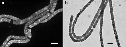
Figure 3.
(a) Beggiatoa filaments hybridized with the universal Bacteria probe EUB338-III (FISH) and counterstained with DAPI, showing regions along the filament that neither hybridized to the FISH probe, nor were stained with DAPI. (b) Light micrograph of Beggiatoa filaments, showing an “empty” region where a cell lysed. Bars, 10 µm

