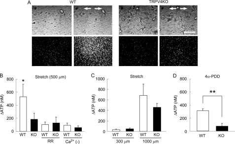
FIGURE 5.
Visualization of stretch-evoked ATP release from primary urothelial cell cultures. A, ATP is robustly released from wild-type (WT) urothelial cells, but poorly released from TRPV4-deficient (TRPV4KO) cells upon stretching. The upper panels show urothelial cells in phase-contrast images, and the lower panels show photon count images (white dots) in the same field. The stretch speed was 400 μm/s, and the distance was 500 μm. Cells were extended transversely (indicated by arrows). Scale bars: 100 μm. B, the average amount of ATP released from TRPV4KO cells in response to mechanical stretch stimulation is significantly smaller than that in WT cells. ATP release is diminished by RR treatment or by the absence of extracellular Ca2+ (Ca2+ (−)) in WT cells. Data presented as means ± S.E. (n > 6). An asterisk indicates a significant difference between WT and the other five groups. *, p < 0.05 (Fisher's protected least significant difference). C, ATP release is minimal at a stretch distance of 300 μm and saturated at 1000 μm in both cell types (also refer to supplemental Fig. S4). Data are presented as means ± S.E. (n = 6). D, 4α-PDD (10 μm) induces robust ATP release by WT cells, but not by TRPV4KO cells. Data are presented as means ± S.E. (n = 6). Significant difference: **, p < 0.005 (Student's t test).

