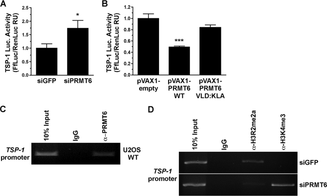
FIGURE 3.
PRMT6 regulates the activity of the TSP-1 promoter by methylating H3R2 in U2OS cells. A, activity of the TSP-1 promoter luciferase (Luc.) reporter gene was assessed in U2OS cells transfected with either siGFP or siPRMT6. The luciferase activity was normalized to Renilla, and the latter was used to assess transfection efficiency. The transfected cells were lysed and assayed for luciferase activity as described under “Experimental Procedures.” The results are expressed as a ratio of firefly luciferase (FfLuc) on Renilla luciferase (RenLuc) fold induction activities. Data represent the means ± S.D. of two independent experiments performed in triplicate. Statistically significant differences as compared with control conditions are as follows: *, p < 0.05; ***, p < 0.001 (Student's t test). B, activity of the TSP-1 promoter luciferase reporter gene was assessed in U2OS cells transfected with either empty vector (pVAX1), wild-type PRMT6 (pVAX1 PRMT6 WT), and catalytically inactive PRMT6 (pVAX1 PRMT6 VLD:KLA). The activity of a TSP-1 promoter luciferase reporter gene was assessed as in A. C, chromatin immunoprecipitation assays were performed with control IgG and α-PRMT6 antibodies. DNA oligonucleotides from the TSP-1 promoter were used in a PCR assay to amplify a DNA fragment. The latter was visualized by ethidium bromide-stained agarose gel electrophoresis. D, analysis of the occupancy of H3R2me2a and H3K4me3 histone marks at the TSP-1 gene promoter by ChIP analysis. ChIP assays were performed with chromatin prepared from U2OS cells transfected or not with control siGFP or siPRMT6. After 72 h, the cells were harvested for ChIP analysis using the indicated antibodies and a primer pair located in the promoter region of TSP-1. ChIPs experiments were performed in duplicate, and the results shown are representative of the duplicates.

