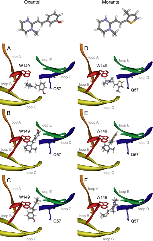
FIGURE 6.
Docking of morantel and oxantel in α 7. Oxantel (left-hand panels: A, orientation 1; B, orientation 2; C, orientation 3) and morantel (right-hand panels: D, orientation 1; E, orientation 2; F, orientation 3) docked into a homology model of the α7 extracellular domain. The molecular structures of morantel and oxantel are shown, with colors indicating C (gray), H (white), N (blue), S (yellow), and O (red). Binding loops are colored orange (loop A), red (loop B), yellow (loop C), blue (loop D), and green (loop E). Residues Trp149 and Gln57 are depicted in red and blue, respectively.

