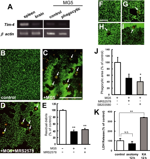
FIGURE 7.
The mechanism of microglial recognition of axon debris appeared to be different from that of apoptotic cells. A, expression profile of Tim-4 mRNA in C57BL/6J mouse spleen, brain, and MG5 cells. The spleen and brain were obtained from a 7-week-old mouse (left panels). The MG5 cells were co-cultured for 0 h (control) or 36 h after transection of the neurites (phagocytic) (right panels). Representative data for RT-PCR for Tim-4 transcripts are shown. B–D, effects of MRS2578, P2Y6 receptor antagonist, on the phagocytosis of degenerating axons by MG5 cells. MRS2578 (10 μm) was added to the culture medium 30 min before transection of the neurites. Double immunostaining of Tuj-1 and ED1 was performed at 36 h after transection of the neurites. MG5 cells engulfed degenerating axons, which were not blocked by MRS2578. Each panel was obtained from the distal part of the severed neurites. Arrows indicate MG5 cells. Scale bar, 200 μm. E, quantitative analysis of the relative number of remaining debris compared with the control (without MG5 cells). Data are represented as mean ± S.E. of three independent experiments. **, p < 0.01 compared with the control. There is no significant difference between MG5 cells and MG5 cells plus MRS2578. F–I, involvement of P2Y6 receptor and p38 MAPK in microglial engulfment of staurosporine-induced apoptotic neurons. The neurons were treated with staurosporine for 48 h. F, control. G, MG5 cells. H, MG5 cells treated with MRS2578. I, MG5 cells treated with SB203580. Scale bar, 50 μm. J, quantitative analysis of the phagocytic area of the data in G–I. K, quantitative analysis of the LDH release in apoptotic cells and degenerating axons. Data are represented as mean ± S.E. of three independent experiments. **, p < 0.01 compared with the control. NS, not significantly different.

