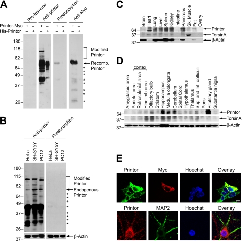
FIGURE 2.
Printor co-distributes with torsinA in multiple tissues and brain regions. A, lysates from transfected SH-SY5Y cells expressing Myc-tagged printor or purified His-tagged printor protein were analyzed by immunoblotting with preimmune serum, anti-printor antibody, anti-printor antibody preabsorbed with recombinant printor protein, or anti-Myc antibody. Recomb., recombinant. B, equal amounts of lysates (50 μg) from the indicated cells were analyzed by immunoblotting using anti-printor, preabsorbed anti-printor, and anti-β-actin antibodies. The asterisks indicate bands that probably represent printor degradation products. C, equal amounts of homogenates (100 μg) from the indicated rat tissues were analyzed by immunoblotting using anti-printor, anti-torsinA, and anti-β-actin antibodies. Sk., skeletal. D, equal amounts of homogenates (100 μg) from the indicated rat brain regions were analyzed by immunoblotting using anti-printor, anti-torsinA, and anti-β-actin antibodies. Sup., superior; Inf., inferior. E, SH-SY5Y cells overexpressing Myc-tagged printor (top) were immunostained with primary antibodies against printor and the Myc tag, followed by detection with secondary antibodies conjugated to Texas Red (Myc, red) or FITC (printor, green). Primary cortical neurons (bottom) were immunostained with primary antibodies against printor and MAP2, followed by detection with secondary antibodies conjugated to Texas Red (printor, red) or FITC (MAP2, green). Hoechst stain was used to visualize the nucleus. Scale bar, 10 μm. All data are representative of at least three independent experiments.

