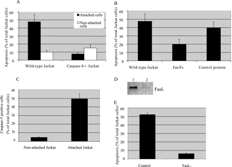
FIGURE 4.
Role of death receptors in Jurkat apoptosis initiated in co-culture with BMSCs. Wild type (WT) and caspase-8-negative Jurkat cells were incubated with a monolayer of BMSCs at a ratio of 1:100 for 48 h (A). Four hours after initiation of co-culture, non-attached Jurkat cells were removed from the co-culture chambers. At the end of co-incubation, all attached cells were trypsinized and labeled with annexin V and anti-CD3 antibody. The percentage of annexin V-positive cells among CD3-positive, attached cells was detected by flow cytometry. Separately, Jurkat cells were co-incubated with BMSCs for 48 h, non-attached cells were removed, and annexin V labeling of non-attached cells was detected by flow cytometry. Fas Fc fusion protein (1 μg/ml) and control Fc protein (1 μg/ml) were added to BMSCs 10 min prior to co-culture initiation (B). Caspase-3 activity of attached Jurkat cells was evaluated using the fluorescent substrate for caspase-3 24 h after co-culture initiation (C). BMSCs were transiently transfected with FasL siRNA or negative control siRNA. Down-regulation of FasL expression by specific siRNA was detected by Western blot (D). Jurkat cells were incubated with a monolayer of wild type and FasL siRNA-transfected BMSCs at a ratio of 1:100 for 48 h. Four hours after initiation of co-culture, the percentage of annexin V-positive cells among CD3-positive, attached cells was detected by microscopy (E).

