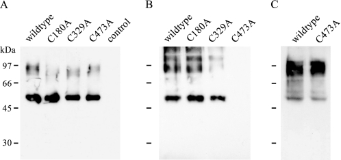
FIGURE 6.
Western blot analysis of cell surface biotinylation of free sulfhydryl groups using maleimide-PEG11-biotin. A, lysates of biotinylated oocytes expressing either wild type or mutant HA-hPAT1. As control, water-injected oocytes were used. B, eluates of neutravidin beads incubated with lysates shown in A. C, eluate of neutravidin beads incubated with lysate of biotinylated oocytes expressing wild type or mutant C473A HA-hPAT1 preincubated for 20 min with 10 mm DTT prior to biotinylation with maleimide-PEG11-biotin. For immunodetection, samples were separated on a 10% SDS gel (8 × 10 cm) followed by incubation with the mouse anti-HA tag antibody.

