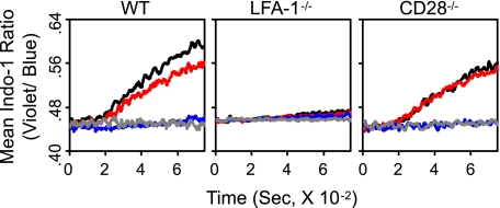
FIGURE 2.
Contributing roles of CD28 and LFA-1 in increase of cytosolic [Ca2+]. Kinetic flow cytometric analyses for changes in cytosolic [Ca2+] were performed with wild type (WT), LFA-1−/−, and CD28−/− 2C T cells being cultured with P1A (1 μm) or QL9 (1 μm) peptide-loaded LdB7-1ICAM-1 pMVs as indicated. 2C T cells were treated with isotype control (black and gray), anti-CD28 (red), or anti-LFA-1 (blue) mAb prior to culture with the pMVs. The flow analyses were initiated 60 s before addition of the pMVs and continued for 720 s afterward.

