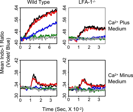
FIGURE 4.
Relationship among levels of pMHC expression, mobilization of intracellular Ca2+, and LFA-1 dependence of extracellular Ca2+ entry. Wild type and LFA-1−/− 2C T cells in Ca2+-containing or Ca2+-free medium were mixed with Ld(high)B7-1ICAM-1 pMVs loaded with P1A (1 μm) (gray) or titrated concentrations of QL9 peptide (black, 200 nm; red, 40 nm; blue, 8 nm; green, 1.6 nm), and changes in cytosolic [Ca2+] were analyzed as in Fig. 2.

