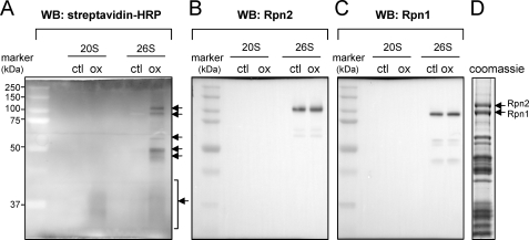
FIGURE 3.
Identification of 19 S regulatory particles that undergo S-glutathionylation after H2O2 exposure. 20 S CP or 26 S proteasomes were incubated with GSH-biotin followed by exposure to H2O2. A, samples were resolved in non-reducing SDS-PAGE gels followed by Western blot analysis with streptavidin-HRP. The presence of GSS-proteasomal subunits is indicated with arrows. The membrane was re-probed with antibodies specific to human Rpn2 or Rpn1 as shown in B and C, respectively. D, subunits of purified 26 S proteasomes were resolved by SDS-PAGE followed by Coomassie staining. The positions of the Rpn1 and Rpn2 regulatory subunits are indicated with arrows.

