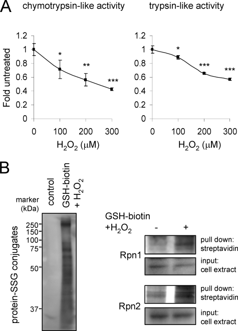
FIGURE 5.
Detection of GSS-Rpn1 and GSS-Rpn2 adduct formation in HEK 293 cells and neutrophils after incubation with H2O2. A, purified human neutrophils were cultured with H2O2 (0–300 μm) for 60 min and then 26 S proteasomal chymotrypsin and trypsin-like activities measured using fluorogenic substrates Means ± S.D. are shown. Each experiment was repeated in triplicate. *, p < 0.05; **, p < 0.01; or ***, p < 0.001. B, left panel shows the increase in total GSS-protein adduct formation obtained from human neutrophils loaded with ethyl ester GSH-biotin and treated with H2O2 for 30 min. GSS-Rpn1 and GSS-Rpn2 levels were detected after pull down with streptavidin-agarose. Representative Western blots are shown. A second experiment provided similar results.

