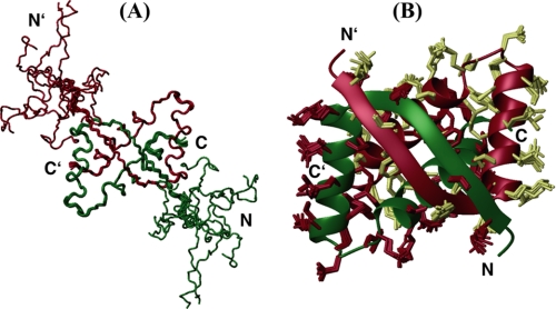
FIGURE 1.
Structure of SvtR. Main-chain trace representation of the structural ensemble of 10 conformers of the full-length protein (residues 1–56) (A) and ribbon representation of residues 11–56 (B). In B, the heavy side chains of the 10 conformers are shown on the ribbon diagram of the lowest energy structure. Monomers are shown in different colors.

