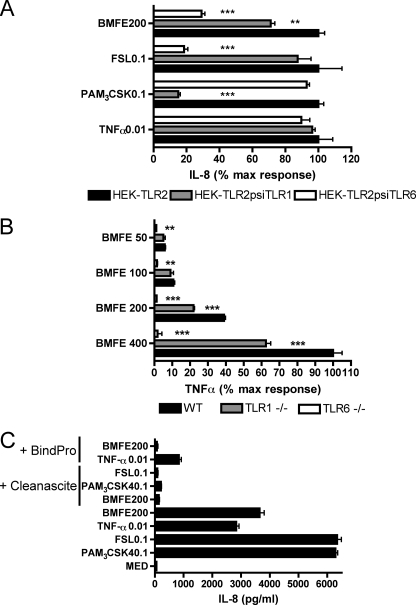
FIGURE 1.
The inflammatory stimuli of BMFE are lipoproteins that primarily signal via TLR2/6. A, HEK-TLR2 cells were transfected with plasmids encoding small interfering (psi) RNA specific for TLR1 or TLR6 before being stimulated with BMFE or control stimuli (doses stated are in micrograms/ml). Accumulations of IL-8 secreted by HEK-psiTLR1 or -psiTLR6 triplicate cultures 20 h post-stimulation are plotted as mean (±1 S.E.) percentages of corresponding HEK-TLR2 responses (mean ± 1 S.E. max IL-8 concentrations as follows: TNFα = 7859 ± 98 pg/ml, PAM3CSK = 9807 ± 175 pg/ml, FSL-1 = 2001 ± 345 pg/ml, and BMFE = 9495 ± 137 pg/ml). Significant differences compared with HEK-TLR2 responses are indicated: ***, p < 0.001; **, p < 0.01. B, peritoneal macrophages from WT, TLR1−/−, TLR6−/− were stimulated with BMFE in triplicate (doses stated are micrograms/ml), and production of TNFα after 20 h is plotted as mean ± 1 S.E. percentages of WT response to 400 μg/ml BMFE (mean ± 1S.E. max TNFα concentration = 188.4 ± 10.36 pg/ml). Significant differences compared with WT are indicated ***, p < 0.001; **, p < 0.01. C, triplicate HEK-TLR2 cultures were stimulated with BMFE or control stimuli (doses stated are micrograms/ml) before or following CleanasciteTM or BindProTM treatment. Data plotted are mean IL-8 ± 1S.E. All data are representative of three independent experiments.

